40 cerebellum labelled
Cerebellum | Description, Anatomy, & Functions | Britannica The Human Brain Like the cerebrum, the cerebellum is divided into two lateral hemispheres, which are connected by a medial part called the vermis. Each of the hemispheres consists of a central core of white matter and a surface cortex of gray matter and is divided into three lobes. Cerebrum Histology - 6 Different Layers with Labeled Diagram Cerebrum of animal's normal brain is composed of paired cerebral hemispheres. In the cerebrum histology you will find the outer gray matter (cerebral cortex) and inner white matter (known as cerebral medulla). Hi there, do you want to learn layers of cerebrum histology with slide images and labeled diagram?
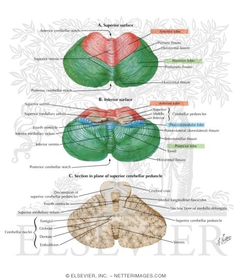
Cerebellum: Gross anatomy and blood supply | Kenhub The cerebellum is a structure that arises from the rhombencephalon or hindbrain. It is located in the posterior cranial fossa inferior to the tentorium cerebelli. It has a superior (tentorial) surface that houses the superior vermis and an inferior (occipital) surface for the inferior vermis.

Cerebellum labelled
Cerebellum: What It Is, Function & Anatomy - Cleveland Clinic The cerebellum, also known as the hindbrain. What is the cerebellum? Your cerebellum is a part of your brain located at the back of your head, just above and behind where your spinal cord connects to your brain itself. The name "cerebellum" comes from Latin and means "little brain." Histological Structure of Cerebellar Cortex - AnatomyLearner Cerebellum histology layers with labeled diagram. Do you want to know more about the cerebellum histology layers? You know cerebellum consists of outer gray matter (cortex) and inner white matter (medulla) in animal. I am going to discuss on cerebellar cortex histology and cerebellar white matter histology separately. BIO EXAM 3 Flashcards | Quizlet The brainstem connects the brain and the: spinal cord. The cerebellum functions in: the planning and coordination of movement. The occipital lobe is labeled __________. C. Name this structure of the cerebellum. arbor vitae. The neurons responsible for hearing, language, memory, and emotions can be found in the __________ lobe.
Cerebellum labelled. Anatomy of the Cerebellum and its Function - ThoughtCo The cerebellum is involved in several functions including: Fine movement coordination. Balance and equilibrium. Muscle tone. Sense of body position. The cerebellum processes information from the brain and peripheral nervous system for balance and body control. Activities such as walking, hitting a ball and playing a video game all involve the ... Cerebellum - W-Radiology The cerebellum is a large structure with two cerebellar hemispheres(4). The hemispheres of the cerebellum are connected by a central part called the cerebellar vermis. The vermis runs along the midsagittal plane and separates the hemispheres vertically. Finely spaced and parallel grooves cover the surface of the cerebellum or the cerebellar cortex. The Cerebellum - Structure - Position - Vasculature - TeachMeAnatomy The cerebellum, which stands for "little brain", is a structure of the central nervous system. It has an important role in motor control, with cerebellar dysfunction often presenting with motor signs. In particular, it is active in the coordination, precision and timing of movements, as well as in motor learning. Cerebellum: histology, layers, cell types and anatomy | Kenhub The cerebellum, located dorsal to the pons and the medulla, is one of the primary structures of the hindbrain. It lies under the occipital and temporal lobes of the cerebral cortex. The cerebellum is an integral structure in transmitting sensory signals to the motor portion of the brain.
Cerebellum | Radiology Key ANATOMY. Grossly, the cerebellum is a small structure situated posterior and inferior to the bulk of the cerebrum (Fig. 16-1). It constitutes approximately 10% of the brain by weight but contains about half as many neurons as the cerebral hemispheres. The three lobes of the cerebellum contain nine lobules, distributed with three lobules in the ... Cerebellum: Functions, Structure, and Location - Simply Psychology The cerebellum, which stands for 'little brain', is a hindbrain structure that controls balance, coordination, movement, and motor skills, and it is thought to be important in processing some types of memory. Cerebellum | Radiology Reference Article | Radiopaedia.org The cerebellum is supplied by three bilateral arteries from the vertebrobasilar system: superior cerebellar artery (SCA): branch of the distal basilar artery anterior inferior cerebellar (AICA): branch of the proximal basilar artery posterior inferior cerebellar (PICA): branch of the distal vertebral arteries Superior cerebellar (SCA) Labeled Brain Model Diagram - Science Trends The midbrain, which is often referred to as the mesencephalon, is the part of the brainstem found highest in the brain. The midbrain connects the other portions of the brainstem to the cerebrum and cerebellum. The midbrain is divided into three different regions: of the tectum, the tegmentum, and the ventral tegmentum.
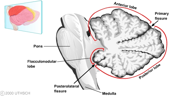
Cerebellum: Function, Anatomy, And Its Location - WebMD Located toward the back of the brain, the cerebellum is one of the densest structures in the brain and is well protected from trauma compared to the brain stem, frontal, and temporal lobes. When... Cerebellum: Definition, Location, and Functions - Verywell Mind The cerebellum is the largest structure of the hindbrain and can be found in the back portion of the skull below the temporal and occipital lobes and behind the brainstem. When looking at the brain, the cerebellum looks much like a smaller structure separate from the brain, found beneath the hemispheres of the cerebral cortex. Anatomy of the cerebellum | Osmosis The cerebellum can be divided into three lobes; the anterior lobe, the posterior lobe, and the flocculonodular lobe. From a superior view, we can identify the anterior lobe, functionally referred to as the spinocerebellum, which is responsible for the regulation of muscle tone and adjusting on-going movements. Labeled imaging anatomy cases | Radiology Reference Article ... This article lists a series of labeled imaging anatomy cases by body region and modality. Brain CT head: non-contrast axial CT head: non-contrast coronal CT head: non-contrast sagittal CT head: non-contrast axial with clinical questions CT ...
The cerebellum and cognition - ScienceDirect
BIO EXAM 3 Flashcards | Quizlet The brainstem connects the brain and the: spinal cord. The cerebellum functions in: the planning and coordination of movement. The occipital lobe is labeled __________. C. Name this structure of the cerebellum. arbor vitae. The neurons responsible for hearing, language, memory, and emotions can be found in the __________ lobe.
2_05 Cerebellum
Histological Structure of Cerebellar Cortex - AnatomyLearner Cerebellum histology layers with labeled diagram. Do you want to know more about the cerebellum histology layers? You know cerebellum consists of outer gray matter (cortex) and inner white matter (medulla) in animal. I am going to discuss on cerebellar cortex histology and cerebellar white matter histology separately.
Cerebellum (Section 3, Chapter 5) Neuroscience Online: An ...
Cerebellum: What It Is, Function & Anatomy - Cleveland Clinic The cerebellum, also known as the hindbrain. What is the cerebellum? Your cerebellum is a part of your brain located at the back of your head, just above and behind where your spinal cord connects to your brain itself. The name "cerebellum" comes from Latin and means "little brain."

Draw a labelled diagram of human brain.

Culmen (cerebellum) - Wikipedia

The human cerebellum with lobules I-X labelled from the ...
draw neat diagram of human brain and label medula and ...

1.184 Otak tengah Gambar, Foto Stok & Vektor | Shutterstock

Learn About Diagram Of Cerebellum | Chegg.com

Cerebellar degeneration - Wikipedia
Anatomy of the cerebellum - Wikipedia

Cerebellum Posterior View Diagram | Quizlet
The Anatomy of the Human Brain

Cerebellum Labeling #1 Diagram | Quizlet

cerebellum - Google Search | Cerebellum anatomy, Human brain ...
Cerebellum: Gross anatomy and blood supply | Kenhub

Sheep brain images | Lab | Amherst College

Cerebellum - an overview | ScienceDirect Topics
Cerebellum: Gross anatomy and blood supply | Kenhub

Draw a labeled structure of the human brain. Write the ...

Cerebellum Function, Location & Anatomy | What is the ...
Labelled Diagram of the Entire Brain-Dorsal View
Cerebellum Vector Art Stock Images | Depositphotos

Cerebellum Diagram | Quizlet
Human Brain labelled stock illustration. Illustration of ...
Cerebellum

Describe the structure of the human brain with a neat diagram

With the help of a labelled diagram of lateral view of ...

Brain Functions Vector Illustration Labeled Explanation Organ ...
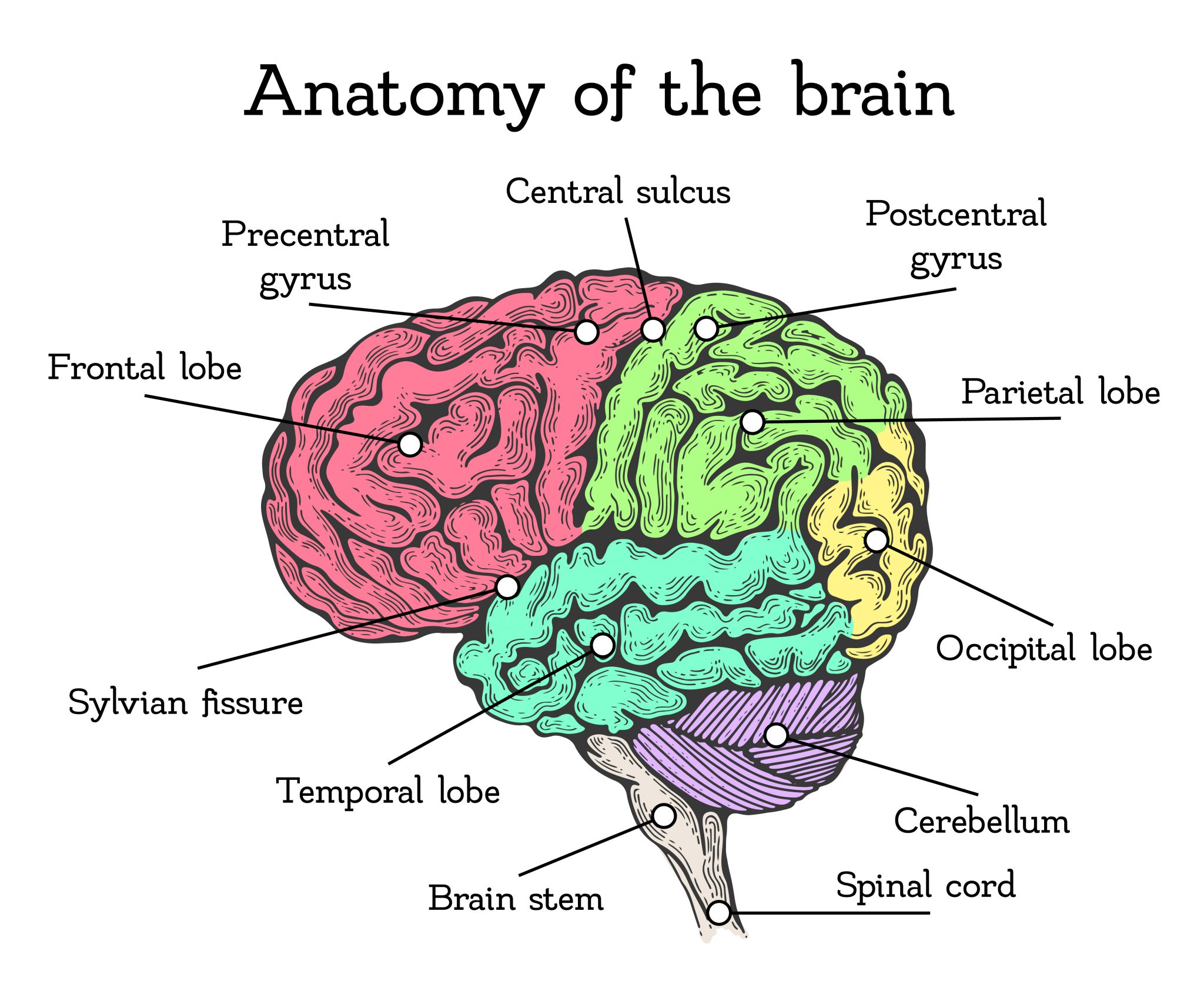
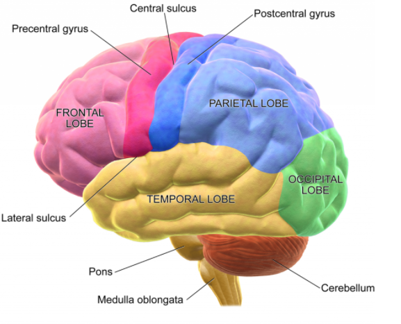
Lobes of the Brain: Cerebral Cortex Anatomy, Function ...
3 Bagian Utama dari Otak Manusia 3 Pound

Brain 1 | anatomyumftm
Draw a neat diagram of the human brain and label any four parts.

Cerebellum labeled Diagram | Quizlet

21 Cerebellum and Brainstem | Neupsy Key

Brain anatomy labelled diagram, Canvas Print | Barewalls ...

Sheep brain images | Lab | Amherst College

Histology of the Cerebrum and Cerebellum – David Fankhauser

Ilustrasi Vektor Cerebellum Diagram Berlabel Medis Dengan ...

Brain Models
Post a Comment for "40 cerebellum labelled"