45 labeled histology slides
Labeling in Histology - Labtag Blog Here, the use of labels designed to fit microscope slides, with a strong resistance to both direct contact and immersion in harsh chemicals and stains is essential. Slide printers are available as well, though durable stain-resistant labels still remain the more reliable choice. Histology: Labelled Slides | SchoolWorkHelper Histology: Labelled Slides Aorta Basophils Cardiac Muscle Cardiac Muscle Longtudinal Cerebellar Cortex Cerebral Cortex Spinal Cord Eosinophils Epiphyseal plate Femoral Artery Femoral Vein Howship's lacunae Jejunum lamina propria Liver Lymph node Lymphocyte Monocyte Spinal cord (silver) Neutrophil Pacinian corpuscle Peripheral Nerve
all histology slide identification tricks | how to identify histology ... all histology slide identification tricks | how to identify histology slide | easy histology vivaFor Buy Anatomy Module Go Through My Website ...

Labeled histology slides
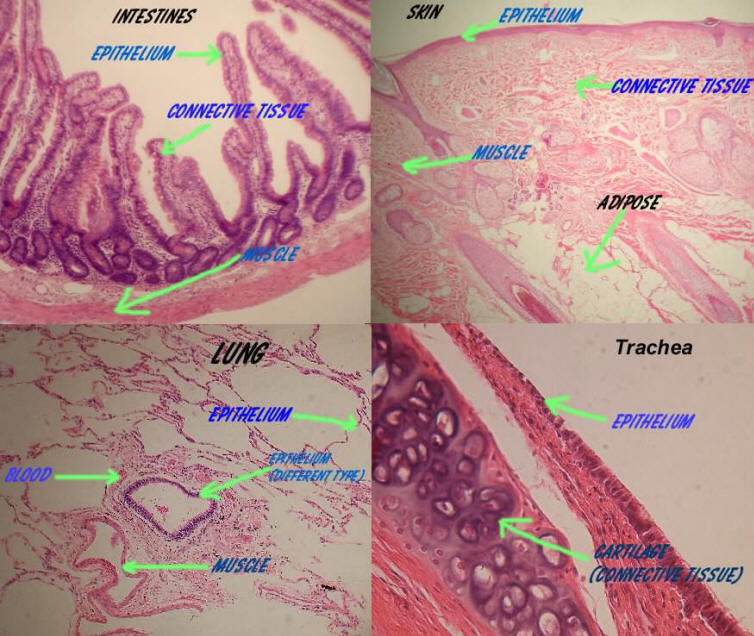
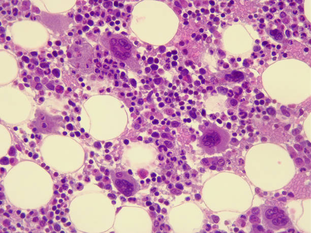
Histology Slides Identification from Different Organ Systems This article will show you histology slides from the following different organs system of an animal's body with identifying features. #1. Histology slide of epithelial tissue #2. General connective tissue histology slide #3. Histology slides of special connective tissue (blood, bone, and cartilage) #4. Muscular tissue histology slide #5. Virtual Slide List | histology - University of Michigan Resources in the University of Michigan Histology Dropbox This collection was originally compiled by Kent Christensen, Ph.D., J. Matthew Velkey, Ph.D., Lloyd M. Stoolman, M.D., Laura Hessler, and Diedra Mosley-Brower. Currently, it is curated by Michael Hortsch, Ph.D. Histology Links | SIU School of Medicine - Siumed.edu The Histology Zoomer ("histology images, tutorials and quizzes", from Yakima Valley Community College) Atlas of Microscopic Anatomy (from Anatomy Atlases, a digital library of anatomy information, curated by Ronald A. Bergman) Human Histology, by Allen L. Bell, University of New England College of Osteopathic Medicine; Virtual Slide Viewing
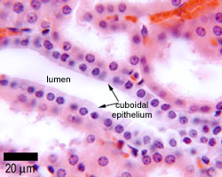
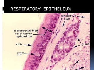
Labeled histology slides. histology human anatomy tissue slides - Quizlet epithelial tissue histology slides. stratified squamous epithelium. simple ciliated columnar epithelium. pseudostratified ciliated columnar epit…. flattened tile-like cells in surface layer, rounder cells in b…. elongated cells, oval shaped nuclei, single layer, projections…. Actually a single layer of cells of varying height, some not r ... 3.1: Examining epithelial tissue under the microscope Sweat glands, salivary glands, mammary glands, adrenal glands, and pituitary glands are examples of glands made of epithelial tissue. Epithelial tissue is often classified according to numbers of layers of cells present, and by the shape of the cells. See Figure 3.1. A simple epithelium is only one layer of cells thick. Slides of Histology | Anatomy and Physiology I - Lumen Learning Slides of Histology Learning Objectives Be able to describe the functions of cells commonly found in connective tissue and identify them. Be able to recognize interstitial (fibrillar) collagens and elastic fibers at the light and electron microscopic levels. Online Histology Made Easy Slides Atlas - MedicForYou W e have prepared an online atlas of histology that has the following histology slides terming it as Histology made easy. These histology slides can be used for practical exams in First Year MBBS while others may find it useful in their ways. Hope the following slides help you in exams and learning it better, though you will have to refer to a histology atlas book.
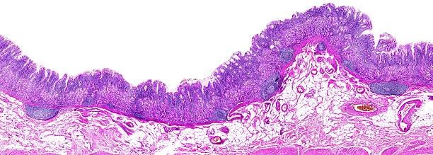
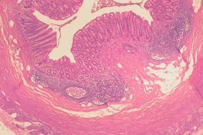
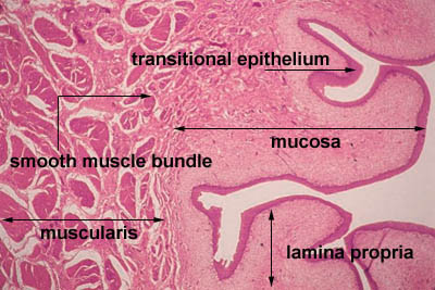
Histology Guide - virtual microscopy laboratory Histology is the study of the microanatomy of cells, tissues, and organs as seen through a microscope. It examines the correlation between structure and function. Histology Guideteaches the visual art of recognizing the structure of cells and tissues and understanding how this is determined by their function. Colon Histology Slide with Labeled Diagram - AnatomyLearner Colon histology layers There are four different tunica layers in the wall of a colon microscope slide - mucosa, submucosa, muscular, and serosa. The structure of the four different layers of the colon microscope slide is almost similar to the structure of a tubular organ. Colon histology slide layers labeled diagram Histology Slides 1 - Loyola University Chicago Histology Slides 1 Slide 1 Mesothelium seen as if looking down on a surface view to see "pavement" effect of the lining cells. Silver stains the intercellular cement dark between adjacent cells. Notice how corrugated the cell membranes are. Mesothelium = the simple squamous epithelium lining body cavities and mesenteries. Slide 2 Virtual histopathology slide box - Knowledge @ AMBOSS The virtual histopathology slide box provides an introduction to the histology of diseased cells and tissues. Each specimen is accompanied by a caption that provides information on staining, magnification, and the structures shown. Virtual microscopy is provided in cooperation with Smart Zoom®. Complementary to this article, the virtual ...
Microscope Slides of Cells and Tissues | Histology Guide This virtual slide box contains 275 microscope slides for the learning histology. Fig 023 Types of Tissue Cells and Tissues Tissues are classified into four basic types: epithelium, connective tissue (includes cartilage, bone and blood), muscle, and nervous tissue. Chapter 1 The Cell Chapter 2 Epithelium Chapter 3 Connective Tissue Chapter 4 Muscle Histology Slides For MBBS 1st Year [With Identification Points] Histology Slides For MBBS 1st Year [With Identification Points] June 25, 2021 by Tauseef Khan In this post, you can view and download all the important Histology slides in the 1st year of MBBS. It includes both general and systemic slides. HistologySlide - Identification of Microscope Slides Histology Slide Identification with Identifying Characteristics Collection of microscope images, labeled diagrams, videos, and more Latest from HistologySlide Slide Identifying Points Slide Identifying Points Slide Identifying Points Slide Identifying Points Slide Identifying Points Slide Identifying Points Slide Identifying Points Histology Slide Identification Tricks | All Histology Slides In 15 ... Medico Darshil Histology Slides Identification Here in this video i tried to describe all histology Slides in 15 minutes. Identifying Epithelium | Review and Practice Questions Anatomy...
Mammal Histology Slide Mikroskop Individual Slide Siap Untuk Peralatan Lab Biologi Versi Bahasa Inggris Label Pada Sisi Geser - Buy Factory Outlet ...
Histology Slides 1 - Loyola University Chicago Welcome to the LUMEN Histology Slide Series. LUMEN
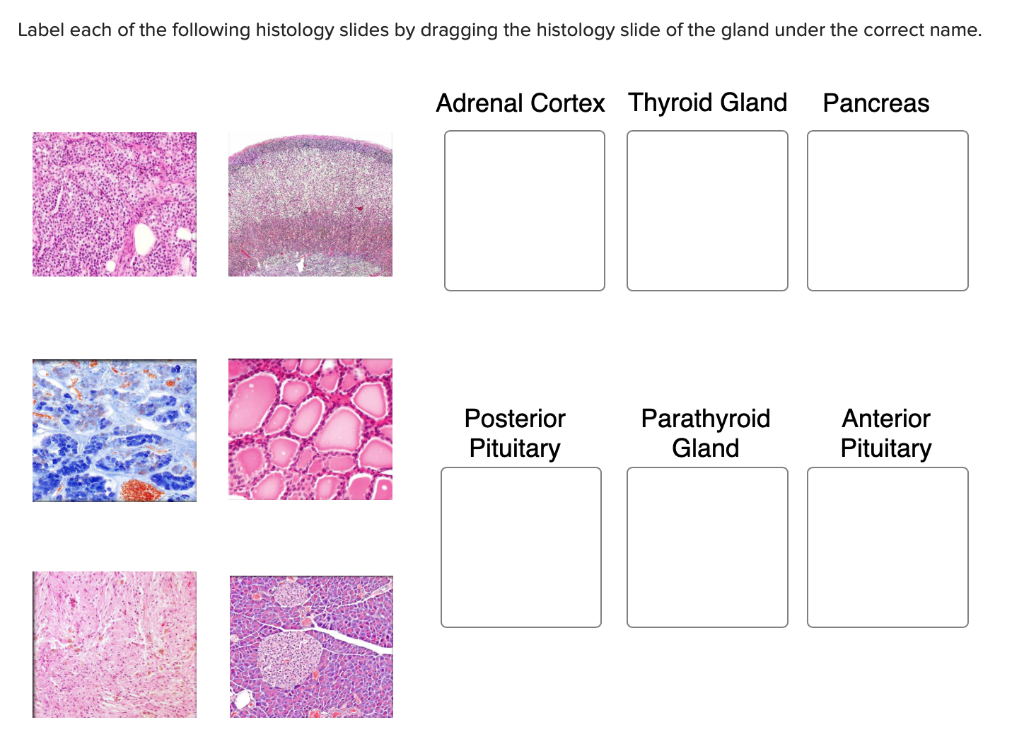
Solved Label each of the following histology slides by ...
Histology Slide Labeling & Preparation - General Data Company, Inc. Our slide labeling solutions are able to withstand the harsh chemicals, reagents and stains of a histology lab's slide staining protocols and process. Each slide is permanently identified and barcodes remain scannable before and after the staining process, as well as through review and archiving. Error-Proof Your Lab's Processes

ovary-3-slide-labelled-histology | SchoolWorkHelper
Histology of the Liver- - SlideShare The Liver • Largest gland of the body. • 1500 grams and 2.5% of total body weight. • Location: - Right hypochondrium - Epigastric region - Left hypochondrium. 3. Vascular Supply of the Liver • Receives dual vascular supply: -Hepatic Portal Vein (75%) - Hepatic Artery (25%) • Both vessels enter the liver via Porta hepatis. 4.

588 Histology Slides Photos and Premium High Res Pictures ...
Learn histology faster With quizzes and flashcards | Kenhub With Kenhub's huge library of histology slides, of course! In our histology atlas, we clearly highlight a given structure on our slides. Comparing several of these slides next to one another is a great way to get a feel for how one tissue differs from another. Enter: our labeled and unlabeled histology tissue identification quiz worksheets.
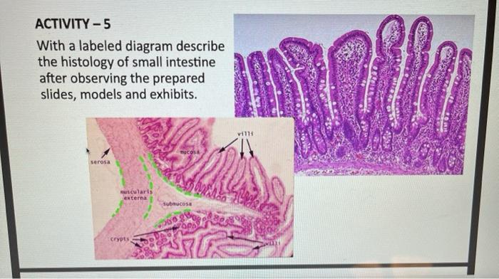
Solved ACTIVITY-5 With a labeled diagram describe the | Chegg.com
Histology Labels | Xylene Resistant - LabTAG Laboratory Labels LabTAG® provides labels and ribbons for every step of the histological process, from tissue extraction and preparation to staining and imaging. Our xylene-resistant labels for histology are designed for microscope slides, paraffin wax blocks, plastic containers, slide boxes stored in deep-freeze conditions, and resin embedding.

Labeled Histology Slides Flashcards | Quizlet
Histology guide: Definition and slides | Kenhub At a histological level, both the heart and blood vessels consist of three layers: Endothelial layer - epithelial tissue formed by simple squamous (endothelial) cells. In the heart, this layer is referred to as endocardium. Muscular layer - smooth muscle in the blood vessels, cardiac muscle (myocardium) in the heart.
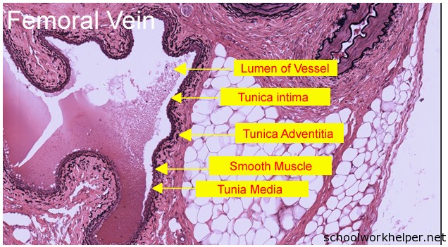
Femoral-Vein-slide-labelled-histology | SchoolWorkHelper
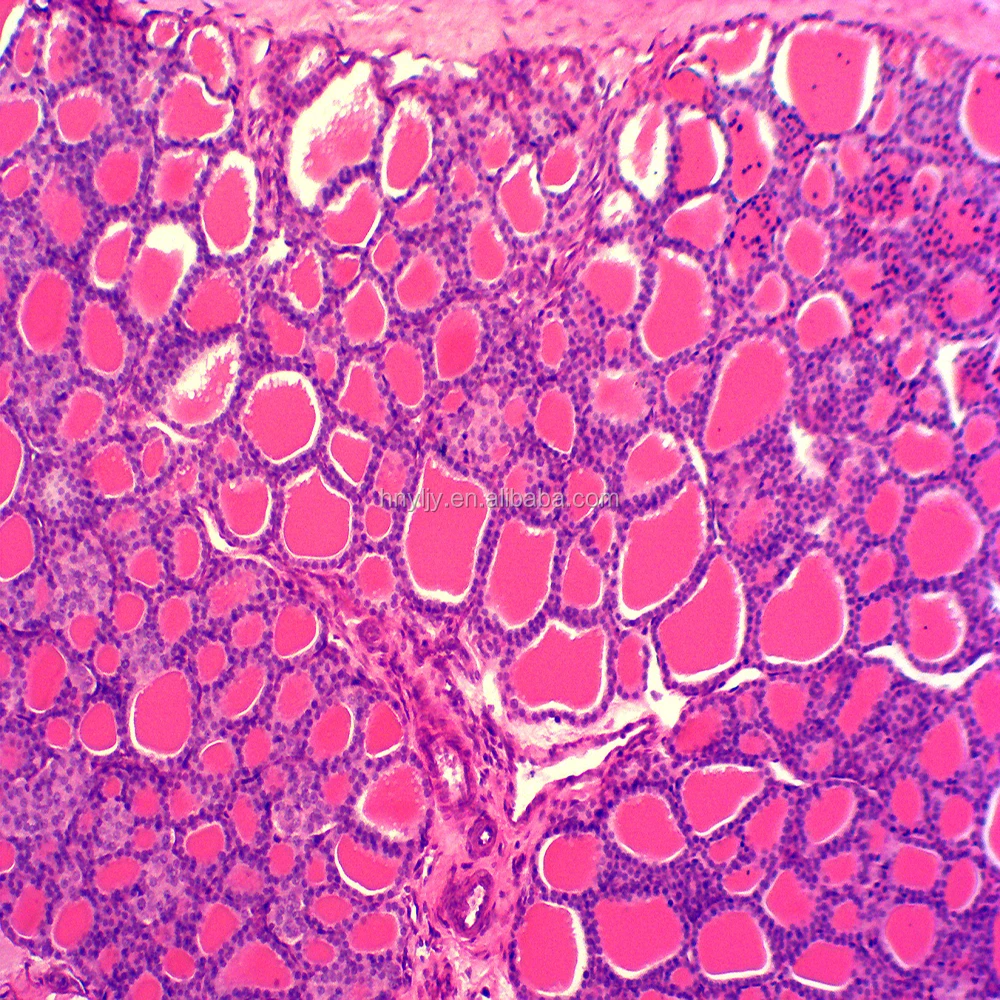
Endocrine System | histology - University of Michigan There are three versions of slide 218 that show a rodent thyroid at three different levels of functional activity: (1) normal slide 218-1 View Image, (2) hypoactivity due to hypophysectomy slide 218-hypo View Image, and (3) hyperactivity slide 218-hyper View Image due to treatment with the drug thiouracil. Compare the tissue shown in each slide ...

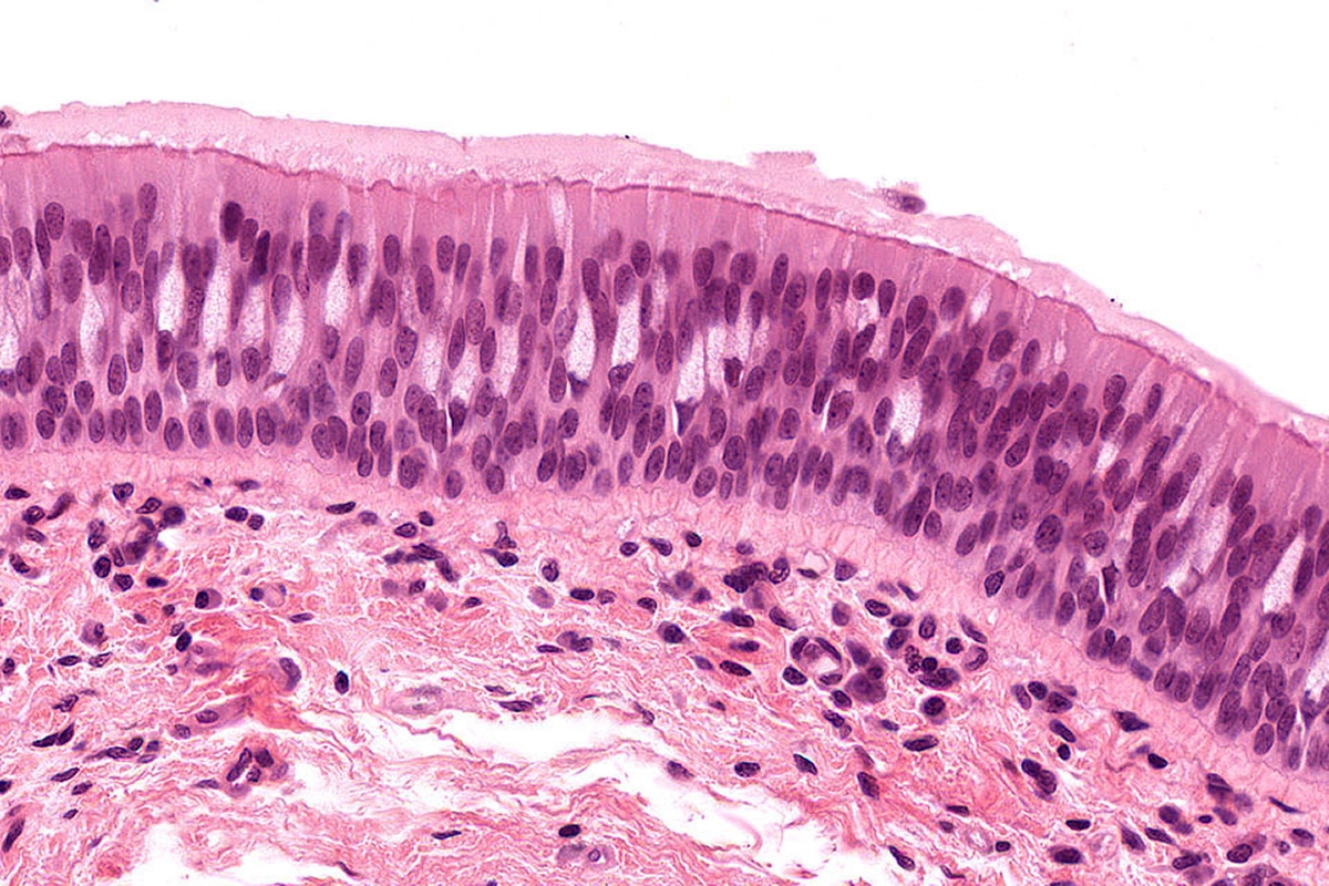
trachea-slide-labelled-histology | SchoolWorkHelper
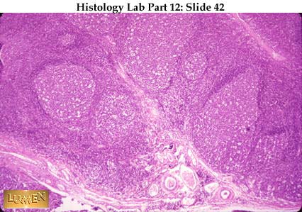
Mbbs part 2 histology slides labelled Mbbs part 2 histology slides labelled. 1. Muhammar Ramzan Ul Reham 1. 3. Apepndix Star shaped leumen Ring of lymph nodules Muhammar Ramzan Ul Reham 3. 5. Colon Deep and closely packed crypts of liberkhun Taenia coli Much more goblet cells Muhammar Ramzan Ul Reham 5.
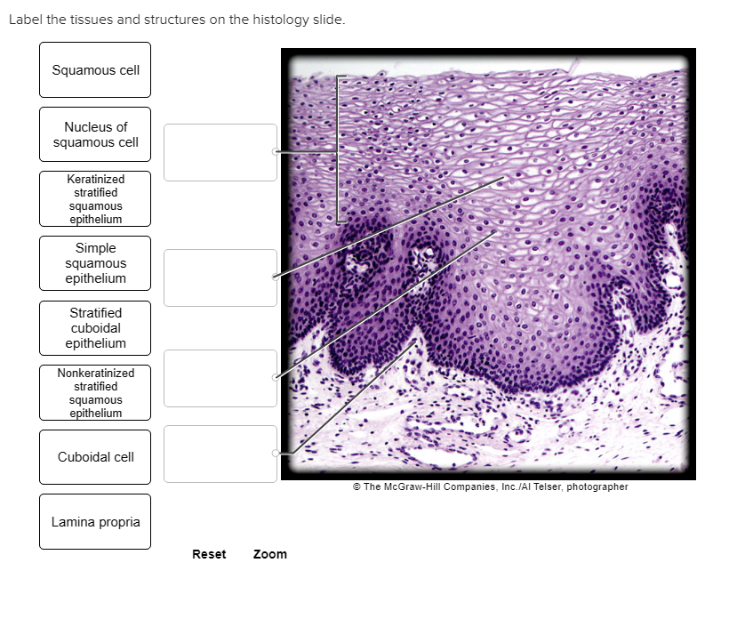
Solved Label the tissues and structures on the histology ...
Histology Microscope Slides | Carolina.com Comprehensive Medical Histology Slide Set Item #311992 $848.00 Connective Tissue Types Microscope Slide Set Item #312034 $88.00 Digestive Tract Microscope Slide Set Item #312106 $65.00 Discovering Epithelial Tissues Self-Study Unit, Microscope Slide Set Item #312008 $91.00 Epithelium Types Microscope Slide Set Item #312016 $73.00
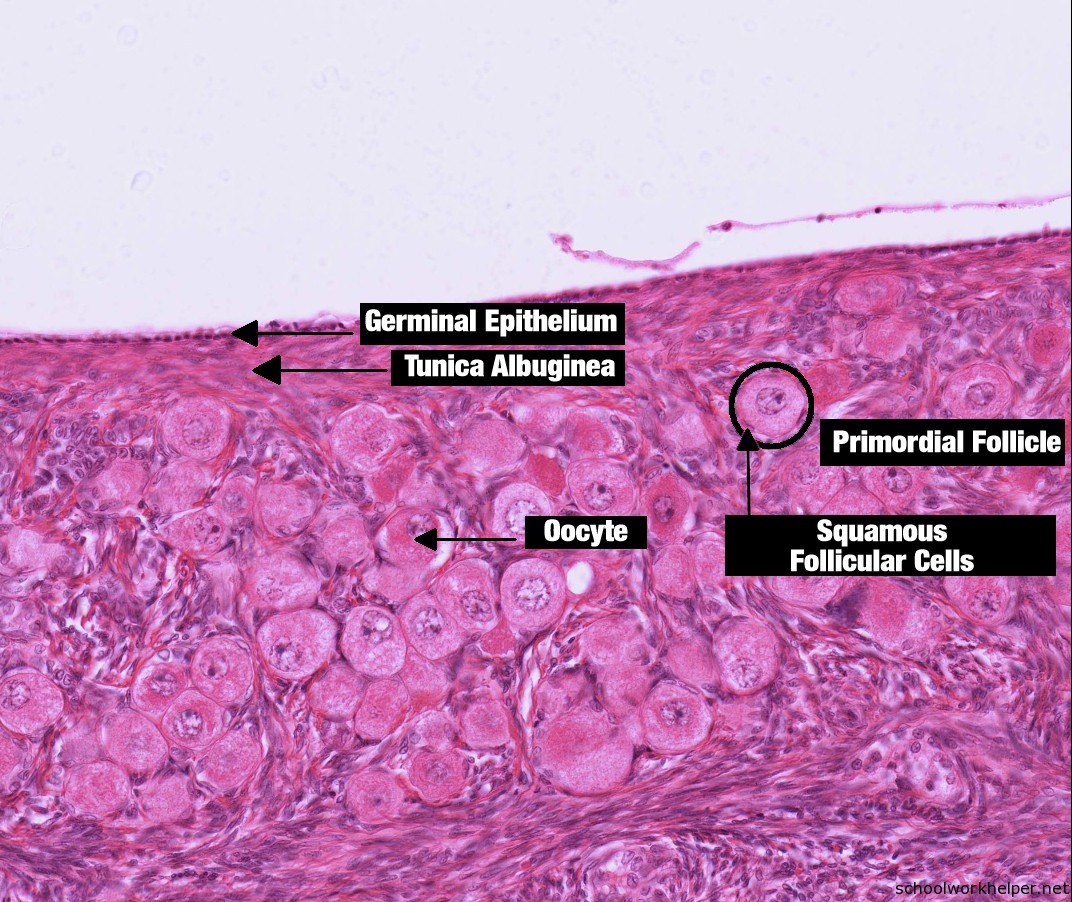
ovary-slide-labelled-histology | SchoolWorkHelper
PDF uniform labeling of blocks and slides in surgical pathology teacing ... consultations), laboratories should label the blocks and slides with at least two patient identifiers, one of which is the patient name. • In the absence of an accession designation or barcode generated by the LIS, blocks and slides that are produced by the laboratory require two OTHER patient identifiers •

Revision of histology slides
Histology Slides - Microscope 50 Histology Human Tissue Slides SKU: OMSK-HH50 In stock 50 professionally made, Prepared Human Tissue slides Educational range of blood, muscle and organ tissue samples Mounted on professional glass slide with sealed cover slips Individually labeled Long lasting hard plastic storage case Recommended for schools and home use You pay: $119.00

Lab.02 Bio.141: Histology (virtual slides) Flashcards | Quizlet
Histology Links | SIU School of Medicine - Siumed.edu The Histology Zoomer ("histology images, tutorials and quizzes", from Yakima Valley Community College) Atlas of Microscopic Anatomy (from Anatomy Atlases, a digital library of anatomy information, curated by Ronald A. Bergman) Human Histology, by Allen L. Bell, University of New England College of Osteopathic Medicine; Virtual Slide Viewing
Atlas of Human Histology
Virtual Slide List | histology - University of Michigan Resources in the University of Michigan Histology Dropbox This collection was originally compiled by Kent Christensen, Ph.D., J. Matthew Velkey, Ph.D., Lloyd M. Stoolman, M.D., Laura Hessler, and Diedra Mosley-Brower. Currently, it is curated by Michael Hortsch, Ph.D.
Histology Slide Jaringan Sistem Saraf Melihat Struktur Sel - Buy Histologi Slides,Slide Mikroskop,Struktur Sel Product on Alibaba.com
Histology Slides Identification from Different Organ Systems This article will show you histology slides from the following different organs system of an animal's body with identifying features. #1. Histology slide of epithelial tissue #2. General connective tissue histology slide #3. Histology slides of special connective tissue (blood, bone, and cartilage) #4. Muscular tissue histology slide #5.
588 Histology Slides Photos and Premium High Res Pictures ...
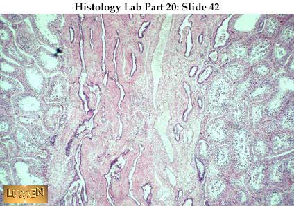
Histology Slides 1
4,500+ Histology Slides Stock Photos, Pictures & Royalty-Free ...

Histology Slides For MBBS 1st Year [With Identification ...
Histology Slides at Thomas Scientific

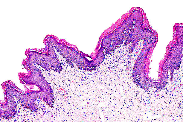
HISTOLOGY, Epithlium Lab, Scalp slide

Teaching medical histology at the University of South ...
Histology Guide - virtual microscopy laboratory

Pathology Slides

femoral-artery-slide-labelled-histology | SchoolWorkHelper
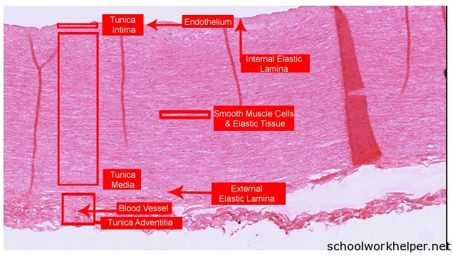
aorta-slide-labelled-histology | SchoolWorkHelper
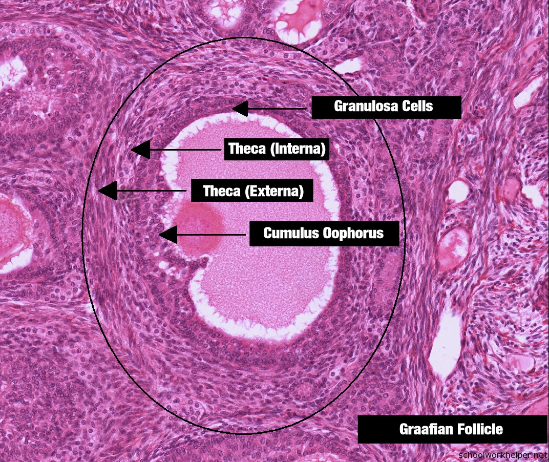
ovary-2-slide-labelled-histology | SchoolWorkHelper
Histology Guide - virtual microscopy laboratory

Images from histology slide preparations from specimen EG 9 ...
Solved] answer asap pleaseeee Label the tissues and ...

A) A histology slide of the entire wound surrounded by the ...
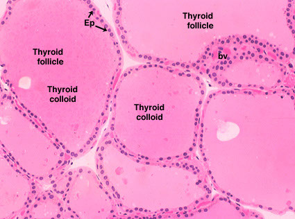
Thyroid Slide
Histology-World! Audio Histology Slide
Epithelia: The Histology Guide
Urethra
4,500+ Histology Slides Stock Photos, Pictures & Royalty-Free ...
Histology of trachea and lung

Pathology Slides
Histology at SIU
Histology Tutor
Atlas of Human Histology
4,500+ Histology Slides Stock Photos, Pictures & Royalty-Free ...
Histology-World! Key Histology Features
Histology Slides 1
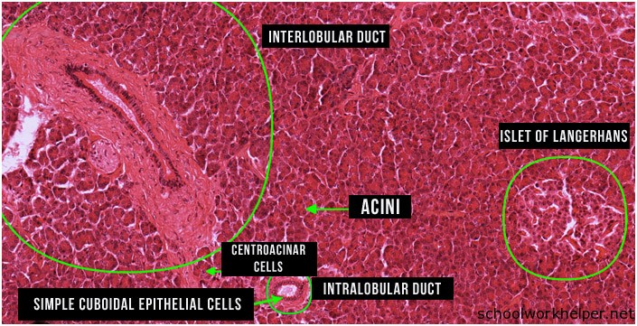
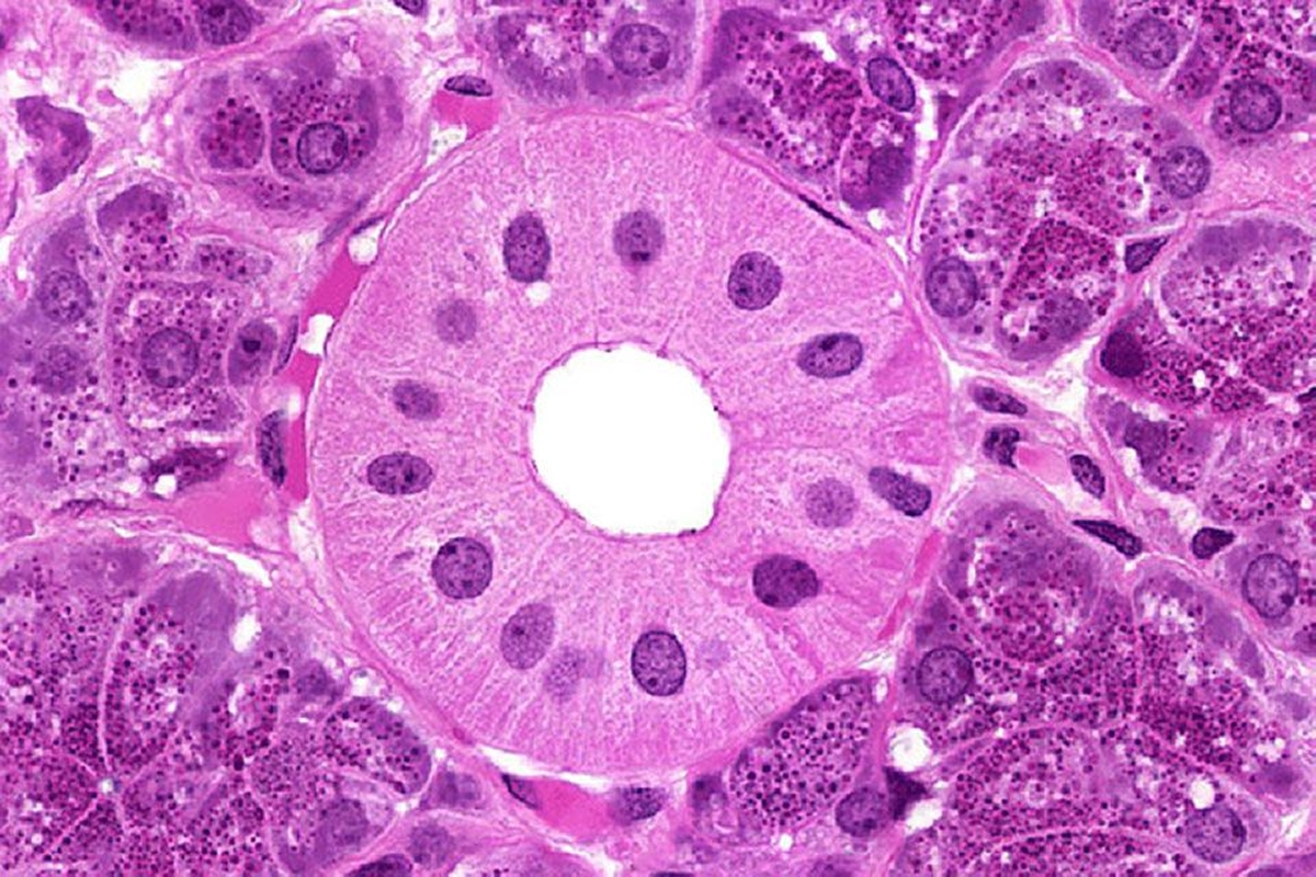
pancreatic-lobule-slide-labelled-histology | SchoolWorkHelper
Histology Microscope Slides | Carolina.com
Post a Comment for "45 labeled histology slides"